
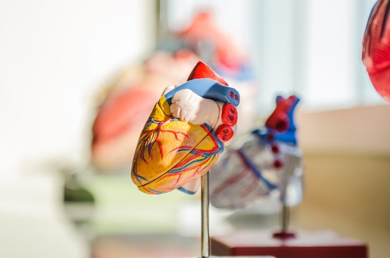
Researchers at Rutgers Robert Wood Johnson Medical School (Rutgers RWJMS) and Robert Wood Johnson University Hospital (RWJUH) have found new biological markers, or indicators, for the early detection of heart disease.
The new, breakthrough ultrasonic imaging technique can identify microscopic changes that occur in heart structure and function.
Go deeper with GlobalData

Discover B2B Marketing That Performs
Combine business intelligence and editorial excellence to reach engaged professionals across 36 leading media platforms.
These changes may help detect early heart disease by leveraging miniaturised ultrasound devices that can be carried in the pocket.
The researchers used artificial intelligence (AI) modelling methods to analyse pixel-based patterns in echocardiogram images and develop interpretations of cardiac conditions that may lead to heart failure.
Using a mouse model of heart failure, they later discovered that these patterns arise from microscopic changes in heart muscle geometry.
Additionally, the researchers established that the new biological markers, or indicators, can help physicians detect cardiac problems earlier and provide important information for helping to plan the appropriate treatment.
US Tariffs are shifting - will you react or anticipate?
Don’t let policy changes catch you off guard. Stay proactive with real-time data and expert analysis.
By GlobalDataRWJMS Cardiovascular Disease and Hypertension Division chief Dr Partho Sengupta said: “By establishing and analysing patterns of pixels obtained from the sample echocardiogram images, we were able to predict presence of heart conditions that can cause heart failure.
“Identifying changes in the heart muscle or cardiovascular function earlier can lead to more proactive interventions and the prevention of serious complications.”
He also said that the new biomarker can be applied to any existing cardiac ultrasound device, such as advanced, miniature hand-held point-of-care ultrasound technology.
Dr Sengupta added: “This has the potential to give more people access to in-depth, expert analysis in a broad range of settings, leading to faster intervention and prevention of serious cardiac disease.”

