
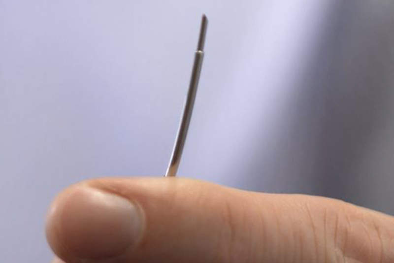
Researchers at Queen Mary University of London (QMUL) and University College London (UCL) in the UK have created a new optical ultrasound needle to enable real-time imaging during keyhole procedures.
The team of surgeons, engineers, physicists and material chemists developed the new technology to address the lack of internal ultrasound imaging feature with existing external ultrasound probes and pre-operative scans.
Go deeper with GlobalData

Discover B2B Marketing That Performs
Combine business intelligence and editorial excellence to reach engaged professionals across 36 leading media platforms.
The technology, which includes a small optical fibre encased in a customised clinical needle, is designed to deliver a brief pulse of light that creates ultrasonic pulses.
A second optical fibre contains a sensor to detect reflections of these ultrasonic pulses from tissue.
As the technology is created to be compatible with current methods such as magnetic resonance imaging (MRI), it could be used in brain or foetal surgery, or with guiding epidural needles.
QMUL consultant cardiologist Dr Malcolm Finlay said: “The optical ultrasound needle is perfect for procedures where there is a small tissue target that is hard to see during keyhole surgery using current methods and, missing, it could have disastrous consequences.
US Tariffs are shifting - will you react or anticipate?
Don’t let policy changes catch you off guard. Stay proactive with real-time data and expert analysis.
By GlobalData“We now have real-time imaging that allows us to differentiate between tissues at a remarkable depth, helping to guide the highest risk moments of these procedures.”
The team designed the needle to be sensitive enough to work during movement inside the body and image centimetre-scale depths of tissues.
During minimally invasive heart surgery in pigs, the technology is reported to have demonstrated a high-resolution view of soft tissues that were up to 2.5cm in front of the device.
The researchers intend to further leverage the technology for other types of minimally invasive surgical methods.

